

For many years, adipose tissue was treated almost as an afterthought. Muscles, fascia, ligaments, tendons and nerves – these were the structures we considered important. Fat? An energy store, thermal insulation, mechanical protection, and not much more. In manual therapy, it has often been regarded simply as a layer we need to pass through to reach the muscle or deep fascia underneath.
Yet research increasingly shows that adipose tissue is far more biologically active than we once thought. It plays a role in metabolic, immune and inflammatory processes, it is innervated and – particularly interesting from our perspective – it can contribute to pain generation.
As a manual therapist, I do not find this particularly surprising. For many years I have been working with subcutaneous tissue that certainly does not behave like a passive layer of fat. Under our fingers, we can find very characteristic and often extremely painful areas of tissue density. These are not isolated points or classic trigger points. They form continuous bands, lines and branches that can often be followed by palpation over considerable distances. Their pathways are remarkably consistent, and pressure applied to one part of the structure can produce not only local pain but also pain perceived elsewhere.
We refer to these structures as lipodynias.
Where did the concept of lipodynia come from?
The term lipodynia
, together with the anatomical identification of these structures, is associated with the work of Dr Radosław Składowski, creator of the FRSc system. They have also been informally described as “octopuses” because larger formations can resemble a central “head” with bands or “tentacles” extending from it.
The term itself is quite straightforward: lipo- refers to fat, while -dynia refers to pain. The terminology was also consulted with a Canadian professor specialising in medical terminology, who confirmed that the term is appropriate for describing painful adipose tissue.
For a long time, however, an obvious question remained. A therapist may feel something under the fingers and the patient may find it painful, but what exactly is there anatomically?
This is where things become particularly interesting.
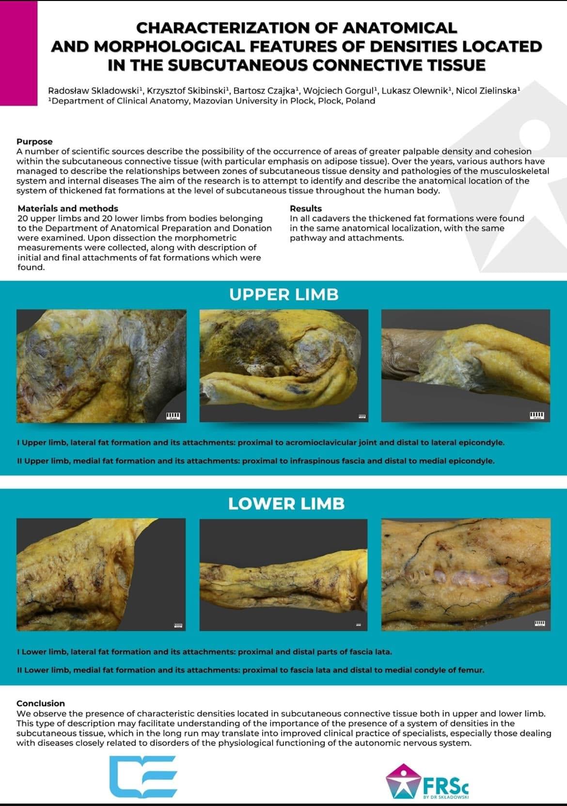
Składowski, together with Krzysztof Skibiński, Bartosz Czajka, Wojciech Gorgul, Łukasz Olewnik and Nicol Zielińska, carried out an anatomical study presented under the title Characterization of Anatomical and Morphological Features of Densities Located in the Subcutaneous Connective Tissue. The work was also presented during a poster session at the IFAA Congress in Gwangju, South Korea.
The researchers dissected 20 upper limbs and 20 lower limbs, performed morphometric measurements, and examined the course and attachment sites of the identified adipose formations.
The results are particularly interesting when compared with what we have been finding through palpation for years. The authors reported that thickened fat formations were found in the same anatomical locations in all examined cadavers, following the same pathways and attachments. That matters.
We are no longer talking only about a therapist's subjective impression that “there is something under my fingers”. These are structures within the subcutaneous tissue that can be anatomically dissected and that demonstrate a repeatable anatomical pattern.
This does not mean that we already understand their complete histology, why they develop or exactly how they may generate pain. What it does tell us is that there are anatomically identifiable structures with repeatable pathways.
And this is where DAT becomes interesting
The story becomes even more intriguing when these anatomical observations are considered alongside current research into subcutaneous adipose tissue itself.
In Human Deep Subcutaneous Adipose Tissue Is Enriched for Inflammatory and Tissue Remodeling Pathways, Yamada and colleagues showed that the fat beneath our skin is not one homogeneous mass. It can be divided into SAT – Superficial Subcutaneous Adipose Tissue – and DAT – Deep Subcutaneous Adipose Tissue. The two are separated by the superficial fascia, fascia superficialis.
More importantly, this is not merely an anatomical division. The two layers are biologically different.
DAT showed greater activity in pathways associated with inflammation, oxidative stress and tissue remodelling. The researchers also found differences in cellular populations, including macrophages and stromal cells.
Does this mean that a lipodynia is a change within DAT?
We don't know.
The Yamada study did not investigate lipodynia or manual therapy, so we cannot simply put an equals sign between the two. But it raises an obvious question. If anatomical dissection reveals repeatable thickened adipose formations, while current research shows that deep subcutaneous adipose tissue is a biologically active environment associated with inflammatory and tissue-remodelling pathways, could at least part of what we call lipodynia be related to DAT or to structures at the interface between these tissue layers?
For me, this is one of the most interesting questions to emerge when we put anatomy, current research and everyday clinical practice side by side.
Adipose tissue really can hurt
There is still the fundamental question: can adipose tissue itself contribute to pain?
Today, the answer is yes.
Lee, Van Dien and Won, in their paper Adipose Tissue as Pain Generator in the Lower Back and Lower Extremity, draw attention to the often overlooked role of adipose tissue in musculoskeletal pain. They describe situations in which adipose tissue may act as a primary pain generator, as well as situations in which it contributes to pain secondarily.
Adipose tissue is innervated and biologically active. Inflammatory processes, structural changes and interactions between adipose tissue, the immune system and nerve endings may all contribute to nociception.
This changes the way we can look at a patient. When something hurts between the skin and the muscle, we do not necessarily have to search for the explanation only in the muscle, joint, ligament or nerve.
Sometimes it is worth stopping exactly where the patient hurts.
Lipodynia and its characteristic pain pattern
Years of clinical observation have shown us something else that is particularly interesting. We associate lipodynias with the autonomic nervous system, particularly sympathetic activity. Within Składowski's clinical concept, three levels of this pattern are described.
The first is non-painful sympathicotonia. The patient does not necessarily experience typical pain at this stage, but we may observe regional hypersensitivity to mechanical stimuli, slower recovery processes within the affected area and the characteristic phenomenon of “moving” or “escaping” trigger points.
The second level is painful sympathicotonia. This is particularly interesting in everyday clinical practice. Pain often becomes worse during inactivity. A patient may go to bed with relatively little discomfort and then wake during the second half of the night with strong, burning or pulling pain. We repeatedly hear patients describe waking between approximately 3:00 and 5:00 a.m. They get out of bed, walk around or start moving, and after some time the pain decreases. They return to bed, fall asleep, and after another period of inactivity the problem may return.
The third level is described by Składowski as severe sympathicotonia progressing towards an algodystrophic presentation. Here, the pain may become constant and severe, much less dependent on either movement or rest, and in some cases it can seriously interfere with sleep.
This classification comes from Składowski's clinical concept and should not be presented as a universally accepted medical classification. The behaviour of the pain itself, however, is certainly something worth paying attention to during clinical history-taking.
What actually hurts within a lipodynia?
Here we move into the area of hypothesis.
Składowski has proposed that one possible source of pain could involve free nerve endings associated with unmyelinated C fibres within the altered subcutaneous tissue. A possible role for mechanoreceptors, including Ruffini endings, has also been considered because of the way these receptors respond to tissue stretch and deformation.
This makes sense as a working hypothesis when compared with what we observe clinically. A precisely directed mechanical stimulus applied along a lipodynia can alter its tenderness and sometimes the patient's symptoms. However, this does not mean that an increased concentration of Ruffini endings or C fibres within lipodynias has already been demonstrated histologically.
That remains a hypothesis and needs to be tested.
This is where I see considerable potential for future research: histology, immunohistochemistry, high-resolution ultrasound, elastography, analysis of innervation and direct comparison between lipodynia tissue and unaffected subcutaneous tissue.
What does this mean in everyday clinical practice?
Perhaps the simplest conclusion is this: we should not automatically push through the subcutaneous tissue in search of the muscle underneath.
It is worth examining the tissue itself. We can assess how the skin and subcutaneous tissue move in relation to deeper layers, compare both sides of the body, assess tissue folding and then follow the tissue longitudinally with our fingers. When we find a clearly altered band, we can examine its continuity, pathway, branches and tenderness.
The patient's response is also valuable information. Does pressure hurt only directly underneath the therapist's finger? Does the sensation travel further? Does palpation reproduce the complaint that brought the patient to the clinic?
These observations can tell us a great deal about a tissue that, until relatively recently, was often largely ignored in manual therapy.
Lipodynias in Pain Relief Techniques
Working with lipodynias is one of the elements of Pain Relief Techniques (PRT). This is not about aggressively “breaking down fat”. It is about learning to recognise altered tissue, identify its course, understand its possible relationship with the patient's symptoms and apply an appropriate therapeutic stimulus.
Within PRT, we use techniques that allow us to work with these densities and bands at different depths and with different levels of intensity. We do not claim that manual therapy “treats DAT”, eliminates inflammation or changes specific cellular populations. We do not yet have evidence to make those claims.
What we do have is extensive clinical experience showing that working with lipodynias can influence pain and function in our patients.
The clinic asked the question. Science is beginning to provide pieces of the answer.
For me, this is perhaps the most interesting part of the whole story.
It began with a clinical observation. Therapists repeatedly found characteristic, painful and reproducible bands within the subcutaneous tissue. Dr Radosław Składowski anatomically identified these structures and introduced the term lipodynia. Anatomical dissections subsequently demonstrated repeatable adipose formations with consistent pathways and attachments.
At the same time, modern research is showing us that adipose tissue can contribute to pain and that superficial and deep subcutaneous adipose tissue are not biologically identical. DAT, in particular, appears to be an interesting biological environment associated with inflammatory and tissue-remodelling pathways.
Is lipodynia a change within DAT? We don't know. Do these characteristic bands have a specific relationship with the autonomic nervous system? We have years of clinical observations and a working hypothesis, but the mechanism still requires further research. Can adipose tissue hurt? Here, the answer is much clearer – yes, adipose tissue can be one of the contributors to pain.
Perhaps it is time, then, to stop treating subcutaneous fat simply as a passive layer that we have to pass through to reach something “more important”.
After more than 35 years of working with patients, I have learned one thing: sometimes the source of a problem needs to be sought far away from where the pain is felt – and sometimes it is located exactly under our fingertips.
References and materials
Składowski R., Skibiński K., Czajka B., Gorgul W., Olewnik Ł., Zielińska N. C
haracterization of Anatomical and Morphological Features of Densities Located in the Subcutaneous Connective Tissue. Anatomical research presented during a poster session at the IFAA Congress, Gwangju, South Korea.
Yamada K. et al. Human Deep Subcutaneous Adipose Tissue Is Enriched for Inflammatory and Tissue Remodeling Pathways. American Journal of Physiology – Cell Physiology, 2025.
Lee S.W., Van Dien C., Won S.J. Adipose Tissue as Pain Generator in the Lower Back and Lower Extremity: Application in Musculoskeletal Medicine.